
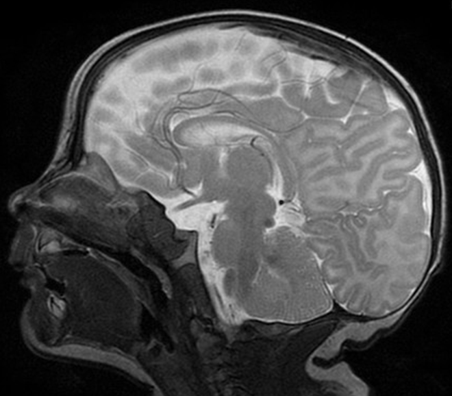
Why is a cranial MRI needed?
Abnormalities of the brain remain quite often undetected by ultrasound and CT examinations, but can be easily diagnosed with MRI. Therewith, the structure and development of the brain can be examined and its response to cerebral injuries (such as haemorrhage or asphyxia) investigated.

Is MRI a safe examination?
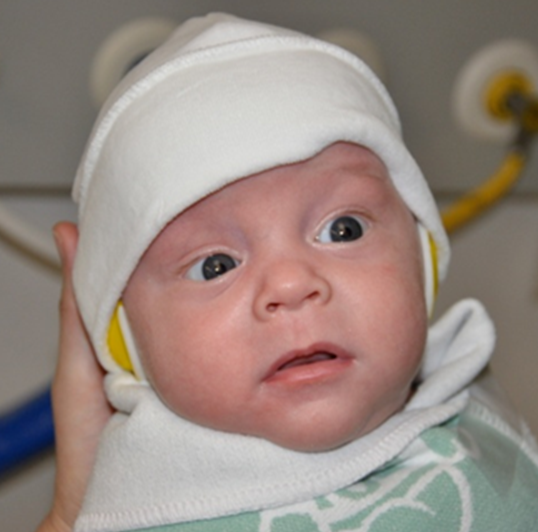
MRI uses a strong magnetic field instead of ionizing radiation to produce cross-sectional images of organs and internal structures in the body. Therefore, no harmful side-effects are known associated with temporary exposure to the magnetic field used by MRI scanners. Because of the quiet noisy imaging process, neonatal noise guards protect the infant's ears in the MRI environment.
How does the examination take place?
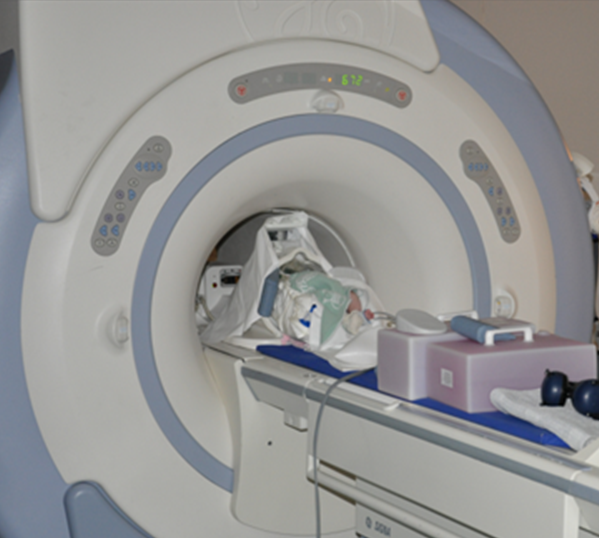
To get the best imaging qualitiy possible, your infant should sleep during the MRI examination. Sedation is only occasionally necessary, because majority of infants can be successfully imaged in natural sleep following a feed and wrapped in a warm blanket. After providing with noise guards, we will wait until the infant has fallen asleep.
Oxygen saturation, heart rate, temperature and, where required, mean arterial blood pressure are monitored throughout the examination, which will not take more than an hour.
How does it go on after the MRI?
After the MRI examination, the neonatologist in charge will inform you about the most important findings. Imaging sequences of the brain especially developed for this study, analysing changes in volume and microstructrue, will only be used for scientific purposes and therefore not communicated separately.